Close Menu
Illustrated Curiosity
    Facebook X (Twitter) Instagram YouTube
    Illustrated Curiosity
    • Earth
    • Economics
    • Environment
      • Climate
      • Environmental Tech
      • Pollution
      • Wildlife
    • Health
      • Health Tech
      • Medicine
      • Nutrition
      • Exercise
    • History
      • Prehistory
      • Ancient History
      • Postclassical Era
      • Modern History
    • Humans
      • Human Brain
      • Psychology
    • Life
      • Animals & Plants
      • Genetics
      • Paleontology
      • Evolution
      • Genetic Engineering
    • Physics
    • Space
      • Astrobiology
      • Astronomy
      • Extrasolar Planets
      • Space Tech
      • Spaceflight
    • Technology
      • Artificial Intelligence
      • Energy
      • Engineering
      • Materials
      • Robotics
      • Vehicles
    Illustrated Curiosity
    Home » Wellcome Image Awards 2017: Finalist show the art of science
    Life

    Wellcome Image Awards 2017: Finalist show the art of science

    March 12, 2017No Comments1 Min Read
    Share
    Facebook Twitter LinkedIn Pinterest Email
    Native to the Pacific Ocean, Hawaiian bobtail squid are nocturnal predators that remain buried under the sand during the day and come out to hunt for shrimp near coral reefs at night. The squid have a light organ on their underside that houses a colony of glowing bacteria called Vibrio fischeri. The squid provide food and shelter for these bacteria in return for their bioluminescence.
    Hawaiian bobtail squid
    Mark R Smith, Macroscopic Solutions

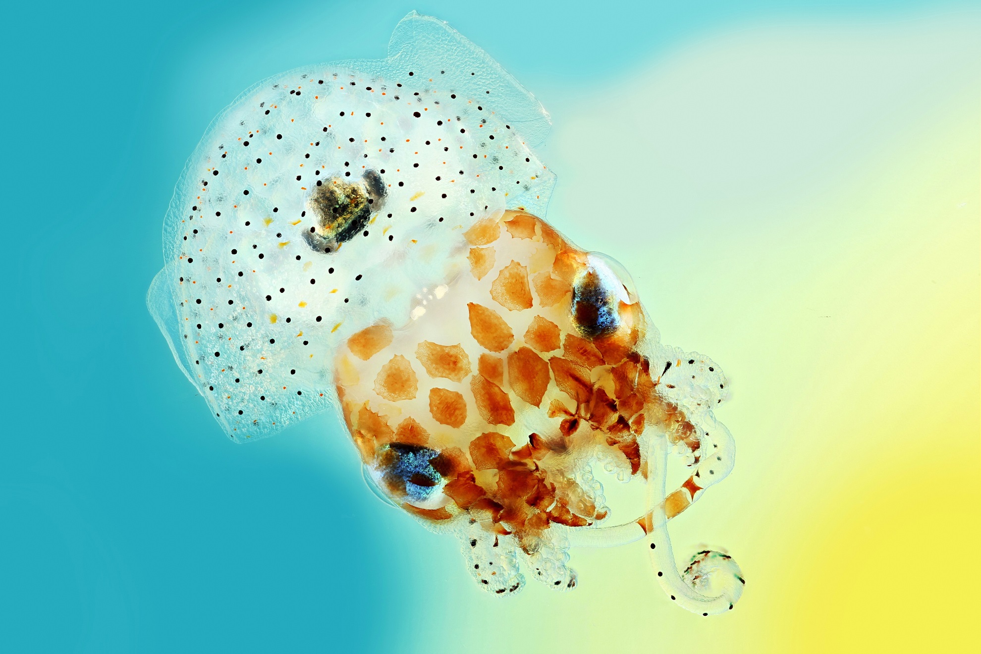
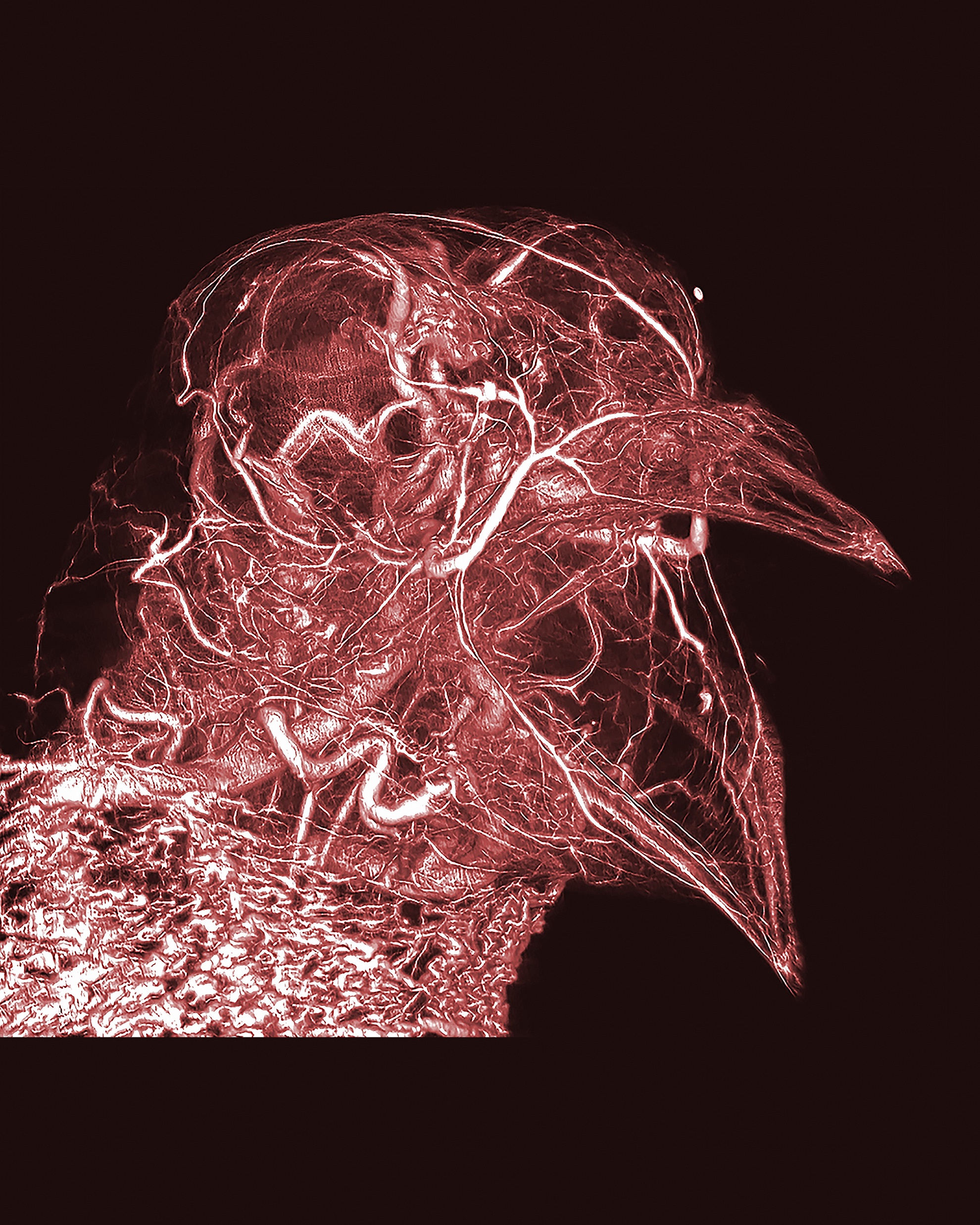
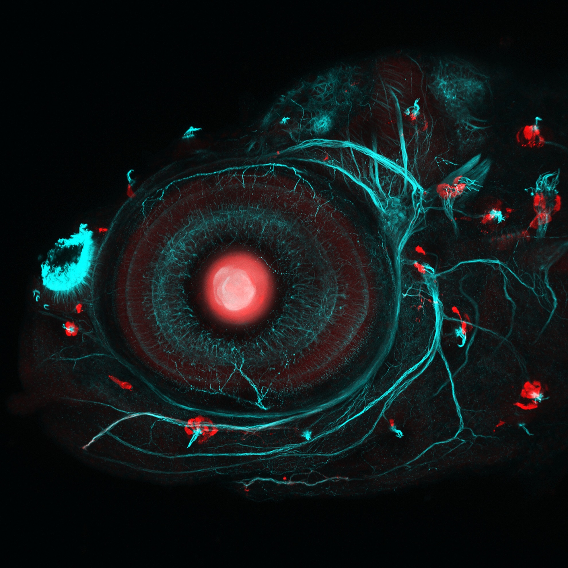
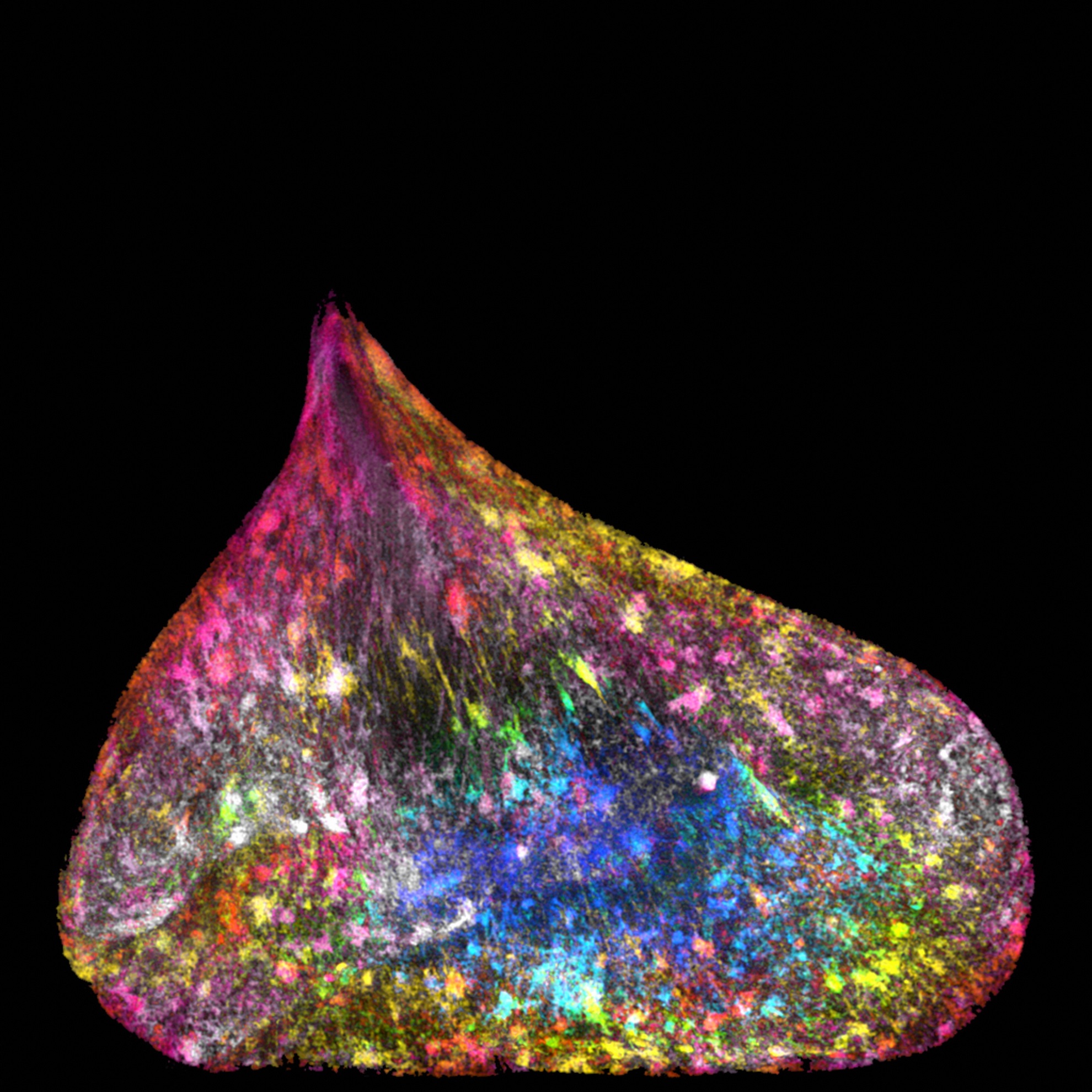
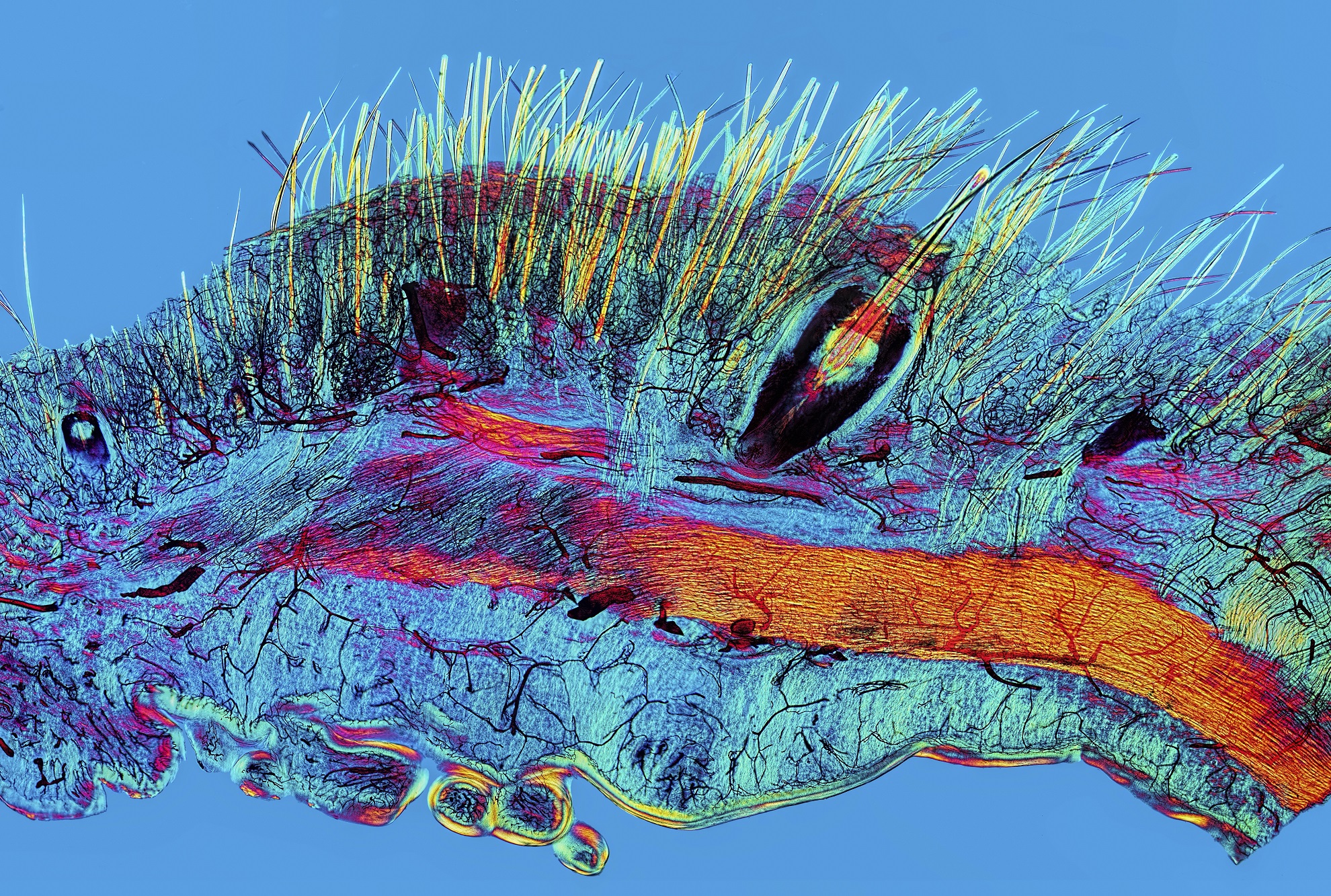
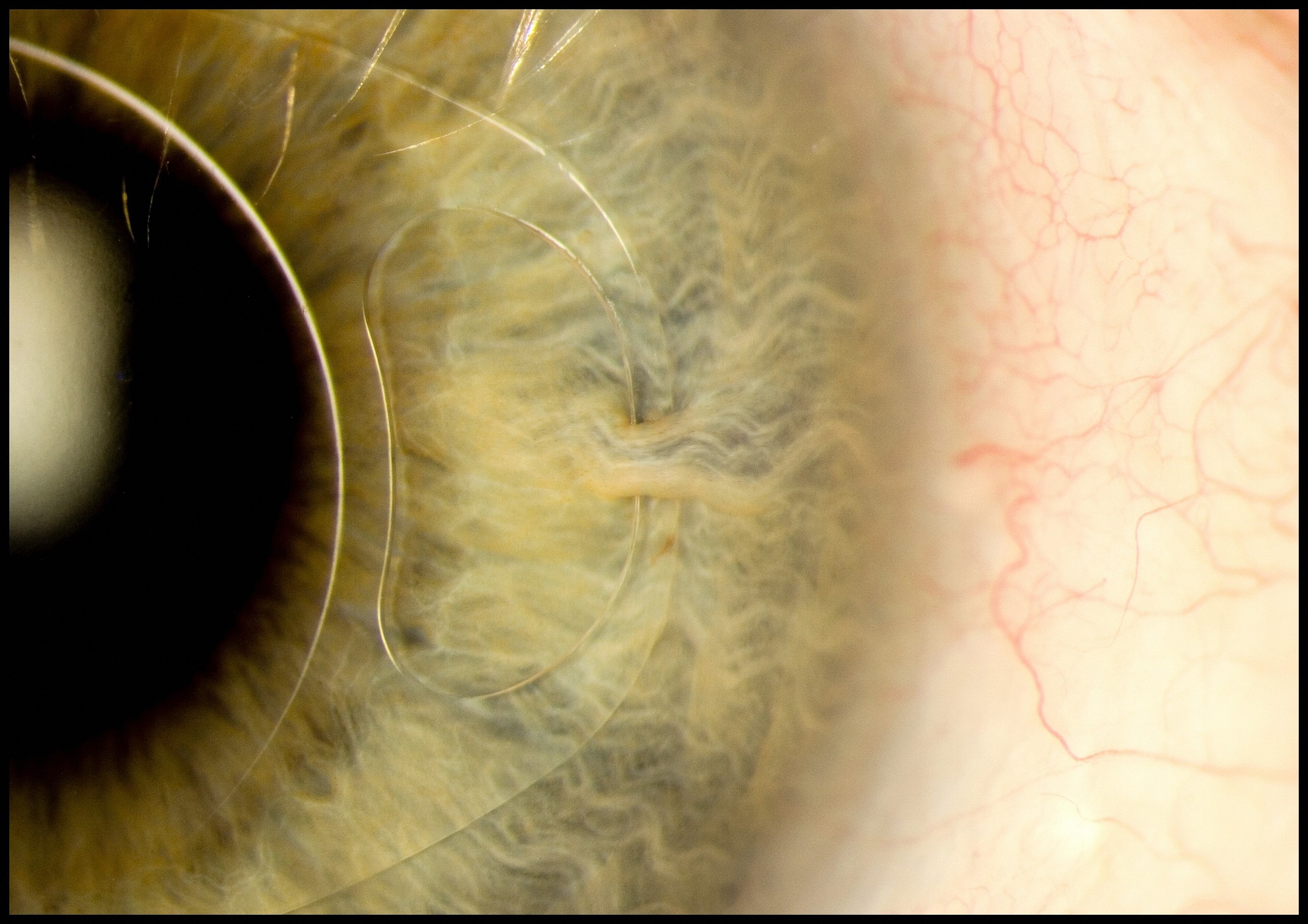
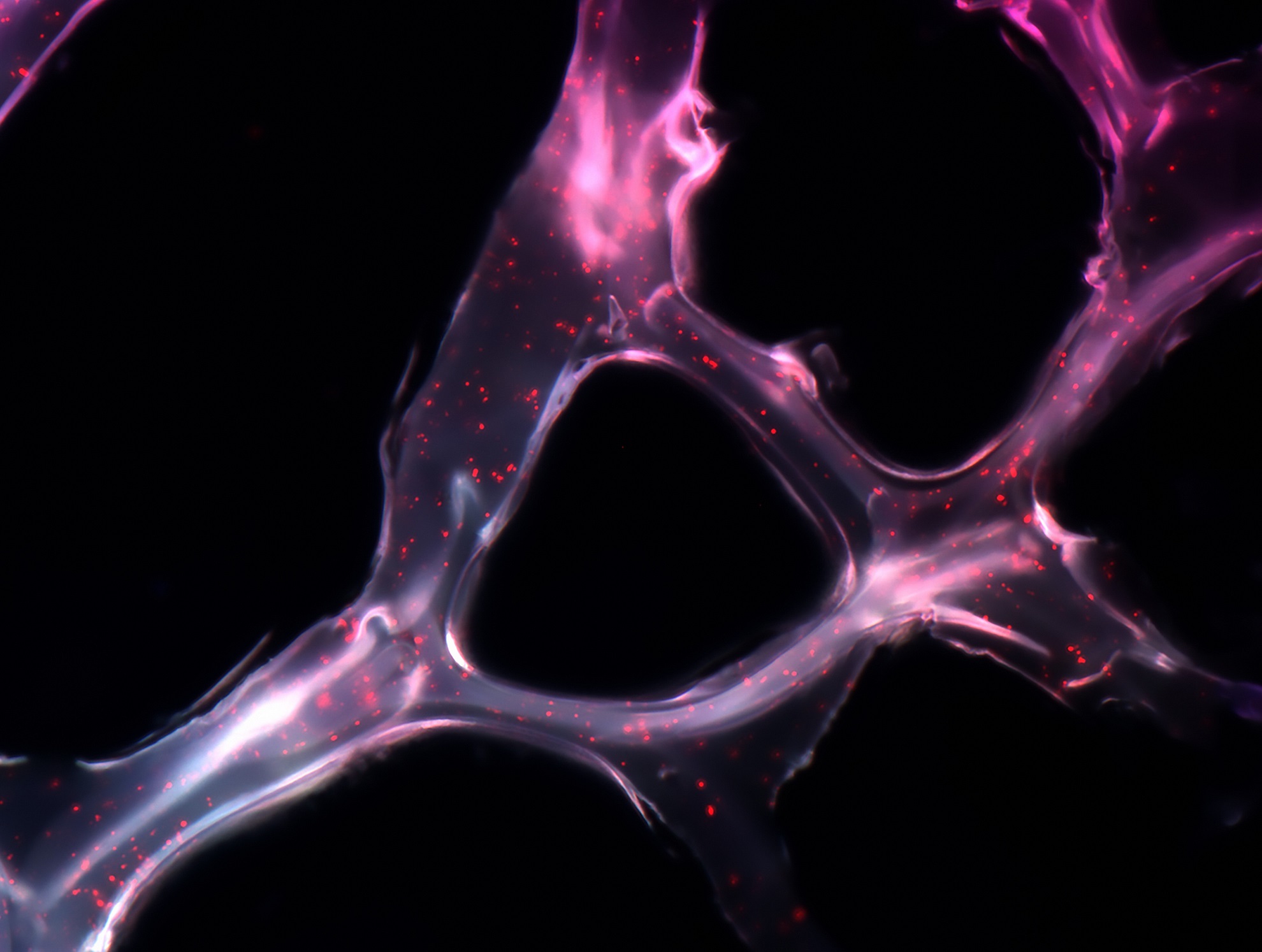
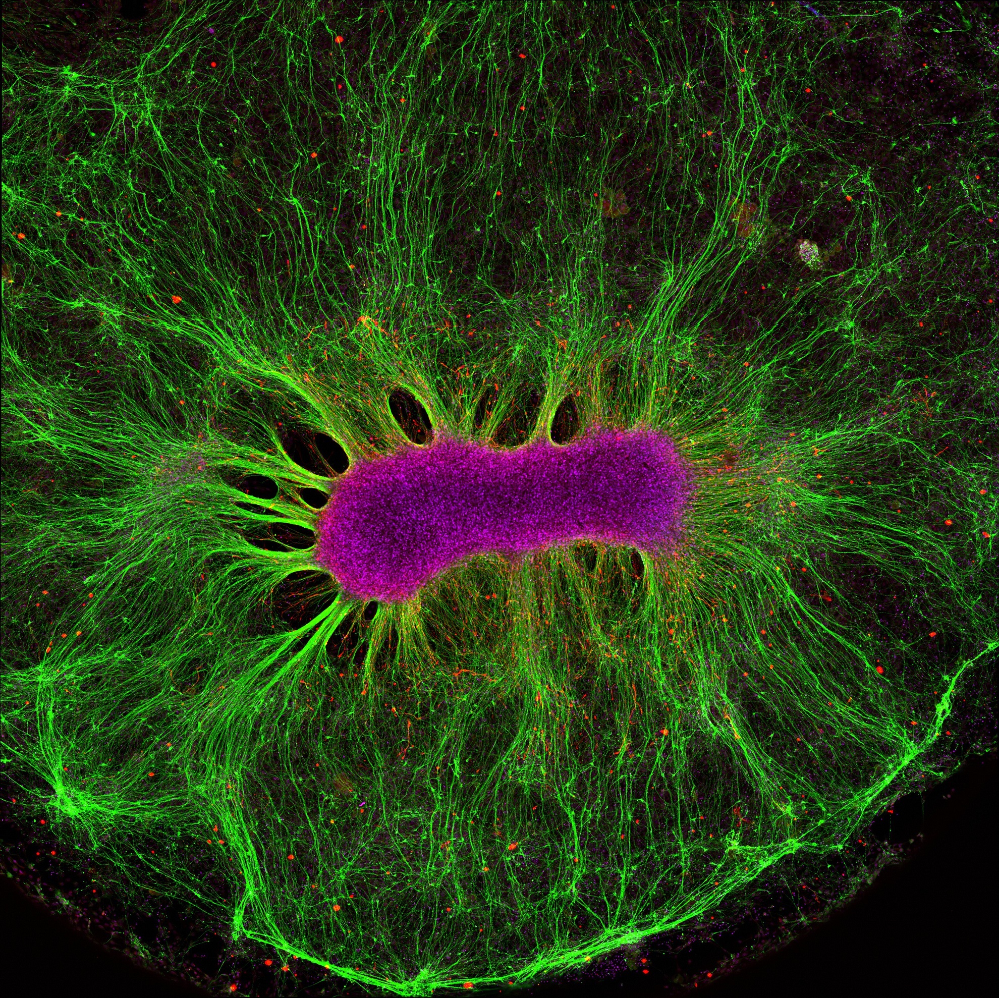
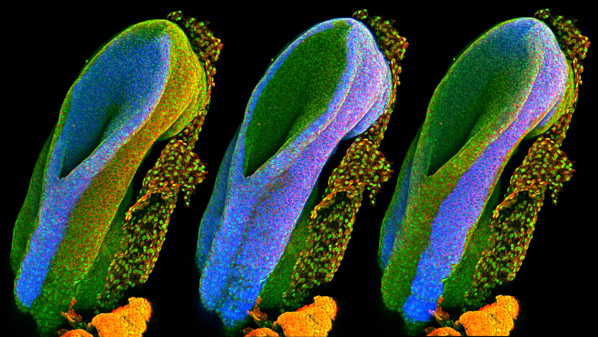
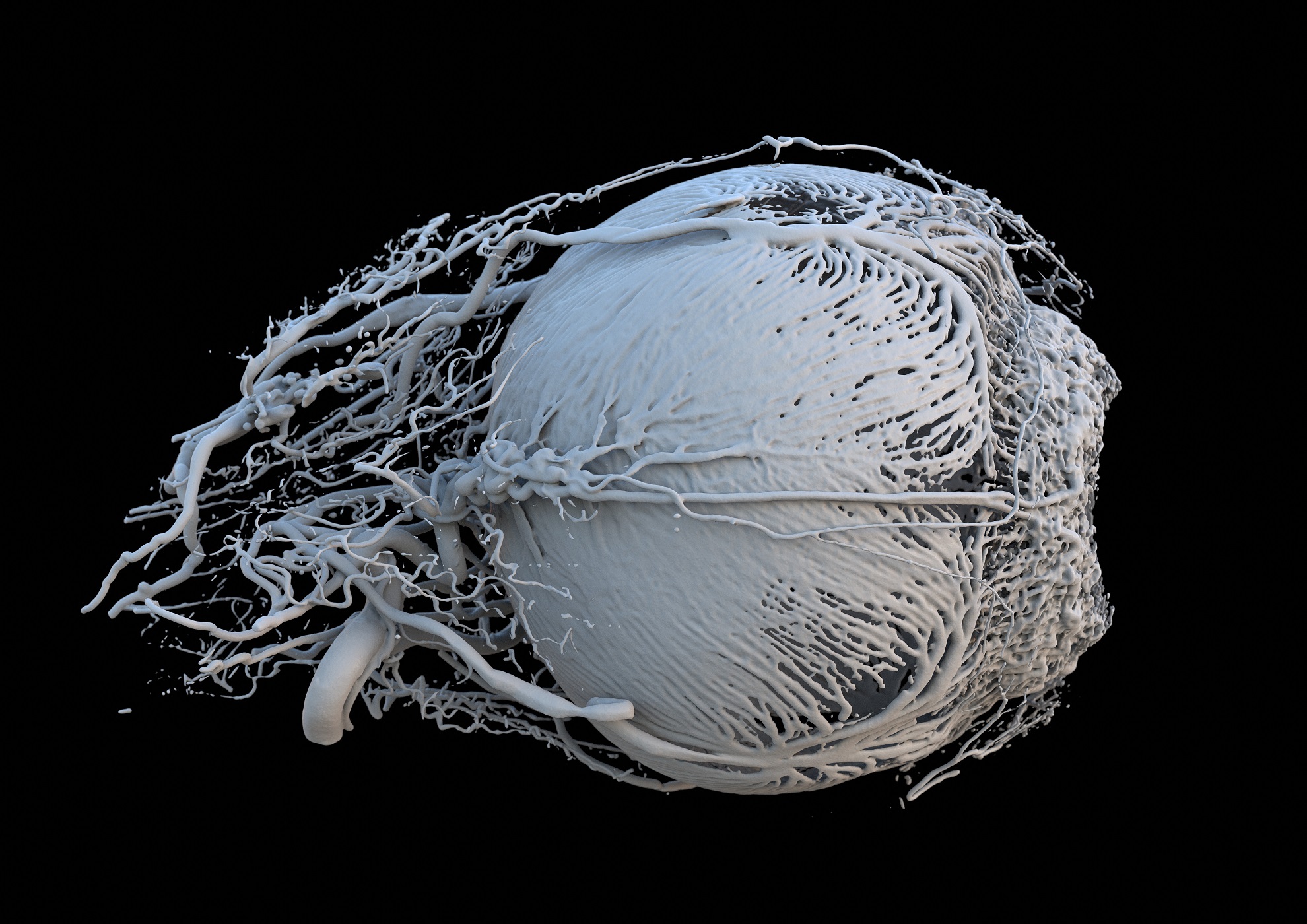
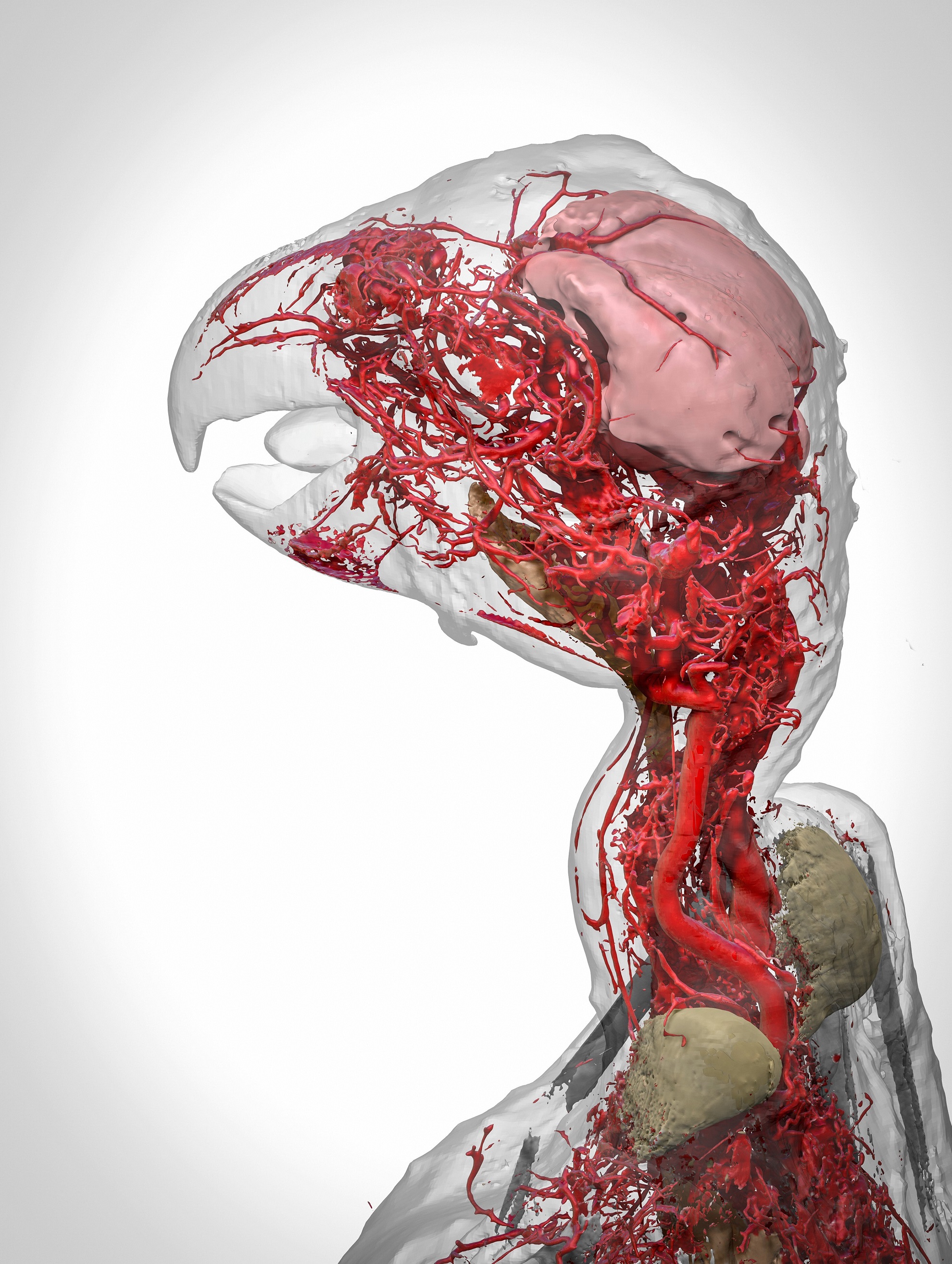
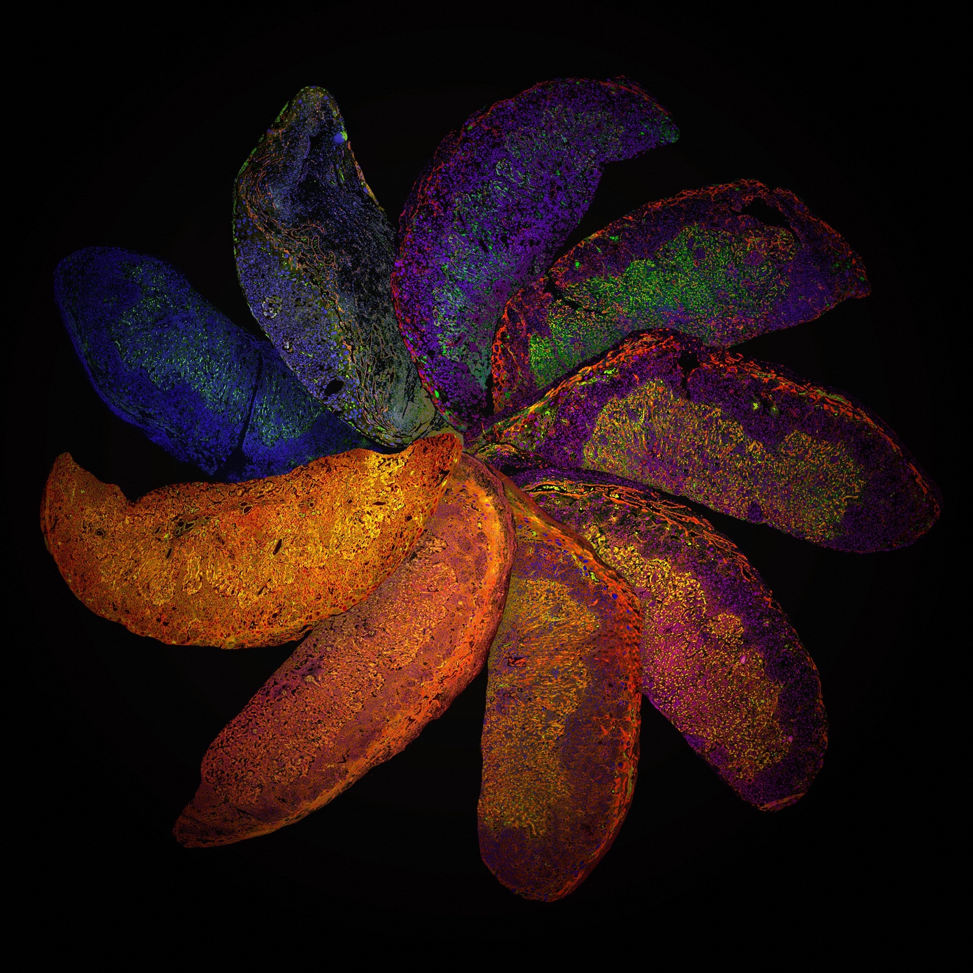
    The finalists of the 2017 Wellcome Image Awards have been announced, showcasing the best science-related imagery from the past year. The winning images will go on display in science centers and public galleries around the world from 16 March 2017.

    The images are judged on quality, technique, visual impact, and their ability to communicate and engage. To achieve the “most eye-catching celebration of science, medicine and life” and to celebrate the scientists, clinicians, photographers and artists who bring science to life through remarkable imaging.

    This image shows a 3D reconstruction of an African grey parrot, post euthanasia. The 3D model details the highly intricate system of blood vessels in the head and neck of the bird and was made possible through the use of a new research contrast agent called BriteVu (invented by Scott Echols). This contrast agent allows researchers to study a subject’s vascular system in incredible detail, right down to the capillary level.
    Pigeon thermoregulation Scott Echols, Scarlet Imaging and the Grey Parrot Anatomy Project
    This four-day-old zebrafish embryo has been modified using two mechanisms – borrowed from the fascinating worlds of bacteria and yeast – that are widely applied in genetics research. A DNA-editing technology called CRISPR/Cas9 was used to insert a gene called Gal4 next to the gene that the researchers wished to study. These Gal4 fish were then bred with special reporter fish to create fish where the gene of interest fluoresces red whenever it is activated.
    Zebrafish eye and neuromasts Ingrid Lekk and Steve Wilson, University College London
    In order for plants and animals to grow and remain healthy, cells need to have the ability to replicate. During cell division, also known as mitosis, the entire DNA content of the cell is copied, with half going to each new cell. DNA is found in a region of the cell called the nucleus, which acts a bit like the brain. This picture shows the nucleus of one of two new daughter cells. The DNA in this cell has somehow become caught, and is being pulled between the two cells. This has caused the DNA to unfold inside the nucleus, and DNA fibres can be seen running through it. As the new cells have moved apart, the tension distributed by the rope-like DNA has deformed the nucleus’s usually circular envelope.
    Unravelled DNA in a human lung cell Ezequiel Miron, University of Oxford
    A polarised light micrograph of a section of cat skin, showing hairs, whiskers and their blood supply. This sample is from a Victorian microscope slide. Blood vessels were injected with a red dye called carmine dye (here appearing black) in order to visualise the capillaries in the tissue, a newly developed technique at the time. This image is a composite made up of 44 individual images stitched together to produce a final image 12 mm in width.
    Cat skin and blood supply David Linstead
    This image shows how an ‘iris clip’, also known as an artificial intraocular lens (IOL), is fitted onto the eye. An iris clip is a small, thin lens made from silicone or acrylic material, and has plastic side supports, called haptics, to hold it in place. An iris clip is fixed to the iris through a 3 mm surgical incision, and is used to treat conditions such as myopia (nearsightedness) and cataracts (cloudiness of the lens). This particular patient, a 70-year-old man, regained almost full vision following his surgery.
    Intraocular lens ‘iris clip’ Mark Bartley, Cambridge University Hospitals NHS Foundation Trust
    Short genetic sequences called microRNAs, which control the proper function and growth of cells, are being investigated by researchers as a possible cancer therapy. However, their potential use is limited by the lack of an efficient system to deliver these microRNAs specifically to cancerous cells. Researchers at MIT have developed such a system, combining two microRNAs with a synthetic polymer to form a stable woven structure a bit like a net. This synthetic net can coat a tumour and deliver the two microRNAs locally to cancer cells.
    MicroRNA scaffold cancer therapy João Conde, Nuria Oliva and Natalie Artzi, Massachusetts Institute of Technology (MIT)
    Neural stem cells have the ability to form all the different cell types found in the nervous system. Here, researchers are investigating how neural stem cells grow on a synthetic gel called PEG. After just two weeks, the stem cells (magenta) produced nerve fibres (green). These fibres grew away from the cell due to chemical gradients in the gel, teaching researchers about how their environment affects their structural organisation.
    Brain-on-a-chip Collin Edington and Iris Lee, Koch Institute at MIT
    Our spines allow us to stand and move, and they protect the spinal cord, which connects all the nerves in our body with our brain. The spinal cord is formed from a structure called the neural tube, which develops during the first month of pregnancy. This series of three images shows the open end of a mouse’s neural tube, with each image highlighting (in blue) one of the three main embryonic tissue types. On the left is the neural tube itself, which develops into the brain, spine and nerves. On the right is the surface ectoderm – the word ‘ectoderm’ comes from the Greek ektos meaning ‘outside’ and derma meaning skin – which will eventually form the skin, teeth and hair. The middle image shows the mesoderm (also from Greek, meaning ‘middle skin’), which will form the organs.
    Developing spinal cord Gabriel Galea, University College London
    A 3D model of a healthy mini-pig eye. The dent on the right-hand side of the image is the pupil, the opening that allows light into the eye. The blood vessels shown are bringing energy and food to the muscles surrounding the iris, which controls the amount of light entering the eye. The smallest vessels seen here are 20–30 micrometres (0.02–0.03 mm) in diameter. The other large vessels are feeder vessels for the retina, the light-sensing region at the back of the eye.
    Vessels of a healthy mini-pig eye Peter M Maloca, OCTlab at the University of Basel and Moorfields Eye Hospital, London; Christian Schwaller; Ruslan Hlushchuk, University of Bern; Sébastien Barré
    This image shows a 3D reconstruction of an African grey parrot, post euthanasia. The 3D model details the highly intricate system of blood vessels in the head and neck of the bird and was made possible through the use of a new research contrast agent called BriteVu (invented by Scott Echols). This contrast agent allows researchers to study a subject’s vascular system in incredible detail, right down to the capillary level.
    Blood vessels of the African grey parrot Scott Birch and Scott Echols
    The Placenta Rainbow highlights differences in mouse placental development that can result from manipulation of the mother’s immune system. These placentas were investigated at day 12 of the 20-day gestation period – the point at which a mouse’s placenta has gained its characteristic shape but is still developing.
    The Placenta Rainbow Suchita Nadkarni, William Harvey Research Institute, Queen Mary University of London
    The brain is composed of two types of matter. Grey matter contains cells, and is responsible for processing information. White matter connects these areas of grey matter, allowing information to be transferred between distant areas of the brain.
    Language pathways of the brain Stephanie J Forkel and Ahmad Beyh, Natbrainlab, King’s College London; Alfonso de Lara Rubio, King’s College London

    Reference:

    Winners’ gallery

    Science
    Share. Facebook Twitter Pinterest LinkedIn Tumblr Email Copy Link

    Related Posts

    Northern Europeans’ Genetic Pre-Historic is Repeated Genocide

    February 6, 2024

    Prehistoric Europeans Consumed Milk Thousands of Years Before Lactose Tolerance

    July 31, 2022

    Kindness To Dogs During Training Matters

    December 28, 2020

    All Dogs Are Descended from the Same Wolf

    November 30, 2020

    The Nobel Prize in Chemistry 2020 Was Awarded to Scientists for Work on Genome Editing

    October 7, 2020

    A Newly Identified Raptor Hunted With The Terrifying Agility of a Cheetah

    June 16, 2020
    Recent Posts
    • How the End of Bretton Woods Reshaped Our Economies — and Our Politics
    • Can the U.S. Really Handle 250% Debt-to-GDP? Why Jackson Hole’s Daring Paper Is Wrong
    • Japan’s Fiscal Trap: What Happens If Austerity Is No Longer an Option?
    • The Rise of Range Extended Electric Vehicles (REEVs): A New Era of Hybrid Mobility
    • The Great Capital Rotation: A Shift from U.S. Treasuries to Gold and Real Assets
    • The Hidden Risks of Short-Term Debt: A Balancing Act in U.S. Fiscal Policy
    • The Looming U.S. Debt Spiral: Understanding the Risks
    • The Looming Recession: Seven Economic Indicators Flashing Red for 2025
    • Europa Clipper: NASA’s Mission to Unlock the Secrets of Jupiter’s Icy Moon
    • Could Hedge Funds Cause Chaos in the U.S. Treasury Market? And What About Inflation?
    © 2025 Illustrated Curiosity

    Type above and press Enter to search. Press Esc to cancel.

    This website uses cookies to improve your experience. We'll assume you're ok with this, but you can opt-out if you wish.Accept Reject Read More
    Privacy & Cookies Policy

    Privacy Overview

    This website uses cookies to improve your experience while you navigate through the website. Out of these, the cookies that are categorized as necessary are stored on your browser as they are essential for the working of basic functionalities of the website. We also use third-party cookies that help us analyze and understand how you use this website. These cookies will be stored in your browser only with your consent. You also have the option to opt-out of these cookies. But opting out of some of these cookies may affect your browsing experience.
    Necessary
    Always Enabled
    Necessary cookies are absolutely essential for the website to function properly. This category only includes cookies that ensures basic functionalities and security features of the website. These cookies do not store any personal information.
    Non-necessary
    Any cookies that may not be particularly necessary for the website to function and is used specifically to collect user personal data via analytics, ads, other embedded contents are termed as non-necessary cookies. It is mandatory to procure user consent prior to running these cookies on your website.
    SAVE & ACCEPT