A new super-resolution microscope will provide new opportunities for medical research.
The microscope is presented in the latest issue of the journal Science. Presented by last year’s Nobel laureate Eric Betzig. The microscope enables us to, among other things, see how different substances are moving about inside a cell.
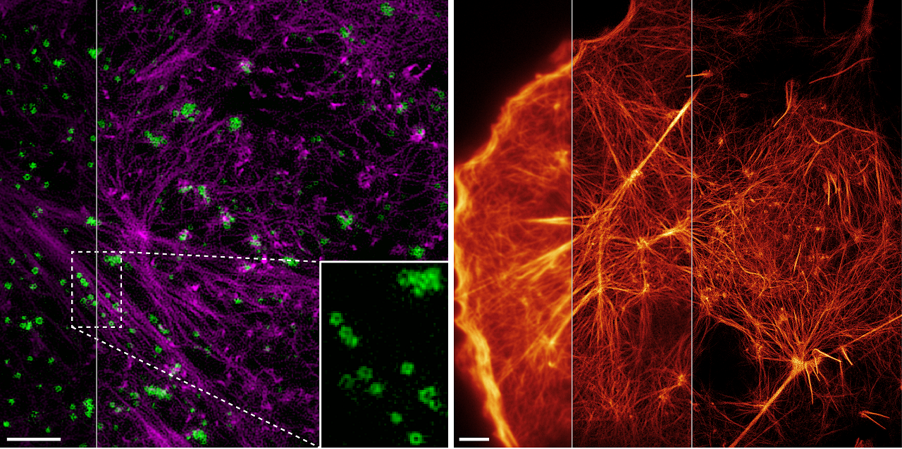
The images of the cells’ inner life were achieved using so-called structured illumination microscopy (SIM microscopy). This SIM technology now enables researchers to make gorgeous 3D movies of living cells at an extreme resolution to reveal what has never been seen before.
Eric Benzig actually received his Nobel Prize for another microscope, but this new SIM microscope is more useful than the one for which he revived his Nobel Prize last year. A major advantage with this new microscope is that it does not harm the cells, as the researchers can now avoid using strong laser light when zooming in on a cell.
The SIM microscope has already led to the new detailed knowledge of what is happening inside our cells, which can be crucial when, for example, we are to develop new pharmaceuticals.
The SIM technology has been in development some time with the Swedish researcher Mats Gustafsson contributing greatly to its development, working on the same team as Eric Betzig before his death in 2011.
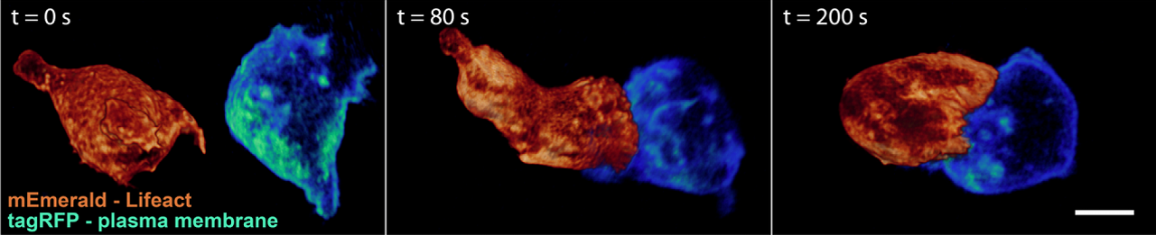
Below a 3D “movie” of how a white blood cell is moving through a matrix of collagen fibers.
_____________
Extended-resolution structured illumination imaging of endocytic and cytoskeletal dynamics
__________________________